GI Lymphoma Panel
Diagnose GI Lymphoma with the use of a simple blood test. When used in conjunction with an ultrasound, avoid the need for biopsy, and get the answers you need.
Gastrointestinal (GI) disease
Differentiating GI Lymphoma from Inflammatory Bowel Disease (IBD) continues to be a challenge in many patients, especially cats. When ultrasound is suggestive and biopsy is not an option, alternative pathways to diagnosis are needed. The VDI Cancer Panel is a test, when used in conjunction with an ultrasound, that can help rule-in LSA in the suspected GI patient. The development of the GI Lymphoma Panel was aimed at improving the detection of small cell LSA and better separating groups for better sensitivity and specificity in the GI differential.
Biomarker Overview
GI Lymphoma Panel is a 4 biomarker panel, 3 of which (along with age) are integrated into the primary diagnostic algorithm: Neoplasia Index®. The markers are:
- Thymidine Kinase Type 1 (TK1): DNA Proliferation Marker
- Haptoglobin (HPT): Inflammatory Marker for cats / C-Reactive Protein (CRP): Inflammatory Marker for dogs.
- Cobalamin (B12): Essential Vitamin affected by and affecting GI IBD/LSA
- Folate: Essential Vitamin involved in DNA synthesis
TK1
TK1, an enzyme responsible for the production of thymidine during cell division, is a proliferation marker of dysregulated replication – a hallmark of cancer. When cell cycles become disrupted/abnormal the TK1 salvage pathway is triggered. In LSA, TK1 values can get exceedingly high.
HPT/CRP
Cancer is an inflammatory disease. When inflammation is present, cancer may be the cause of that inflammation. The major systemic marker of inflammation in cats is Haptoglobin (HPT) and C-Reactive Protein (CRP) in dogs.
B12
Cobalamin is an essential vitamin that plays a number of crucial roles – among them, DNA synthesis. When B12 deficiency exists, cell cycles are incomplete/abnormal resulting in the activation of the TK1 salvage pathway, up-regulating TK1 expression.
Why use the GI Lymphoma Panel?
- Application specific Neoplasia Index® resulting in better Sensitivity/Specificity, especially with Small Cell LSA.
- Indicates if LSA is more likely: Large Cell, Small Cell, or Thoracic.
- B12/Folate report provides additional information on status of the GI tract.
- In absence, or in place of biopsy, the GI Lymphoma Panel can provide a diagnosis.
- Detailed lab reports [see reports below] give you insight to disease and GI status.
Considerations
The GI Lymphoma Panel utilizes disease activity markers that respond to changes in disease state. Use of anti-inflammatory medications like NSAIDS/Corticosteroids and Chemotherapy may impact results, however this does not eliminate the utility of the panel.
Contact VDI for further information regarding the impact of medication on results. All VDI Cancer Panels can be used to monitor the effectiveness of therapy and disease progression.
REFERENCES – Body of Evidence

Boyé, Pierre, et al. “Evaluation of Serum Thymidine Kinase 1 Activity as a Biomarker for Treatment Effectiveness and Prediction of Relapse in Dogs With non‐Hodgkin Lymphoma.” Journal of Veterinary Internal Medicine, vol. 33, no. 4, Wiley, May 2019, pp. 1728–39. https://doi.org/10.1111/jvim.15513.
Euler, Henrik von, Anders B. Öhrvik, et al. “A Non-radiometric Method for Measuring Serum Thymidine Kinase Activity in Malignant Lymphoma in Dogs.” Research in Veterinary Science, vol. 80, no. 1, Feb. 2006, pp. 17–24. https://doi.org/10.1016/j.rvsc.2005.05.001.
Euler, Henrik von. “Monitoring Therapy in Canine Malignant Lymphoma and Leukemia With Serum Thymidine Kinase 1 Activity – Evaluation of a New, Fully Automated Non-radiometric Assay.” International Journal of Oncology, Jan. 1992, https://doi.org/10.3892/ijo_00000175.
Euler, Henrik von, Roland Einarsson, et al. “Serum Thymidine Kinase Activity in Dogs With Malignant Lymphoma: A Potent Marker for Prognosis and Monitoring the Disease.” Journal of Veterinary Internal Medicine, vol. 18, no. 5, Aug. 2004, p. 696. https://doi.org/10.1892/0891-6640(2004)18.
Grobman, M., et al. “Serum Thymidine Kinase 1, Canine-C-Reactive Protein, Haptoglobin, and Vitamin D Concentrations in Dogs With Immune-Mediated Hemolytic Anemia, Thrombocytopenia, and Polyarthropathy.” Journal of Veterinary Internal Medicine, vol. 31, no. 5, Wiley, Aug. 2017, pp. 1430–40. https://doi.org/10.1111/jvim.14787.
Larsdotter, S., et al. “Serum Thymidine Kinase Activity in Clinically Healthy and Diseased Horses: A Potential Marker for Lymphoma.” The Veterinary Journal, vol. 205, no. 2, Elsevier BV, Aug. 2015, pp. 313–16. https://doi.org/10.1016/j.tvjl.2015.01.019.
Meachem, Melissa D., et al. “A Comparative Proteomic Study of Plasma in Feline Pancreatitis and Pancreatic Carcinoma Using 2-dimensional Gel Electrophoresis to Identify Diagnostic Biomarkers: A Pilot Study.” Canadian Journal of Veterinary Research-revue Canadienne De Recherche Veterinaire, vol. 79, no. 3, June 2015, pp. 184–89.
NAKAMURA, Noriko, et al. “Plasma Thymidine Kinase Activity in Dogs With Lymphoma and Leukemia.” Journal of Veterinary Medical Science, vol. 59, no. 10, Japanese Society of Veterinary Science, 1997, pp. 957–60. https://doi.org/10.1292/jvms.59.957.
Selting, K. A., et al. “Serum Thymidine Kinase 1 and C‐reactive Protein as Biomarkers for Screening Clinically Healthy Dogs for Occult Disease.” Veterinary and Comparative Oncology, vol. 13, no. 4, Wiley, July 2013, pp. 373–84. https://doi.org/10.1111/vco.12052.
Selting, K.A., et al. “Thymidine Kinase Type 1 and C‐Reactive Protein Concentrations in Dogs With Spontaneously Occurring Cancer.” Journal of Veterinary Internal Medicine, vol. 30, no. 4, Wiley, May 2016, pp. 1159–66. https://doi.org/10.1111/jvim.13954.
Taylor, Samantha S., et al. “Serum Thymidine Kinase Activity in Clinically Healthy and Diseased Cats: A Potential Biomarker for Lymphoma.” Journal of Feline Medicine and Surgery, vol. 15, no. 2, SAGE Publications, Oct. 2012, pp. 142–47. https://doi.org/10.1177/1098612×12463928.
Thamm, D. H., et al. “Elevated Serum Thymidine Kinase Activity in Canine Splenic Hemangiosarcoma*.” Veterinary and Comparative Oncology, vol. 10, no. 4, Wiley, Oct. 2011, pp. 292–302. https://doi.org/10.1111/j.1476-5829.2011.00298.x.
The Neoplasia Index®: NI GILSA
The Neoplasia Index® is the primary test value used in the diagnostic workup of the GI patient
Neoplasia Index® (NI fGILSA)
The Neoplasia Index® (NI) is meant for the diagnostic rule-in of cancer. Developed as a general cancer marker used in the VDI Cancer Panel, the NI is the primary value used for differential diagnosis of cancer. The NI is unit-less index from 0-10 with 0 being a perfectly normal animal, to 10 being a perfect score for cancer.
Combining independent markers into a unified algorithm improves performance. The base Neoplasia Index® incorporates Thymidine Kinase Type 1 and Haptoglobin/C-Reactive Protein for general cancer rule-in.
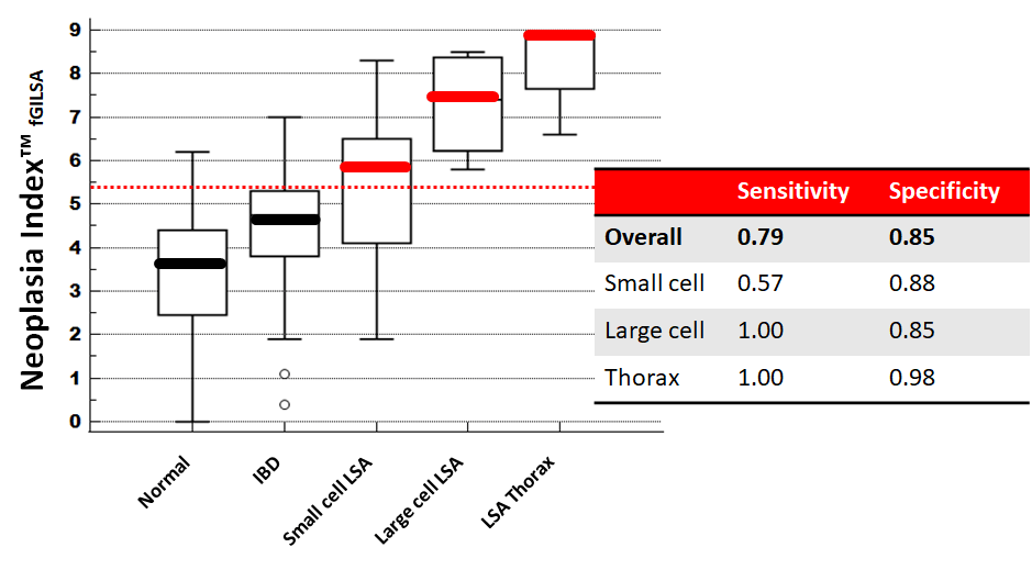
The expanded NI GILSA, is a specific application for GI lymphoma in cats and dogs. It incorporates TK1, HPT/CRP, B12, and Age. As you can see below, in cats, the NI fGILSA separates groups remarkably well. The NI GILSA compensates for B12 deficiency and is able to differentiate small cell vs large cell.
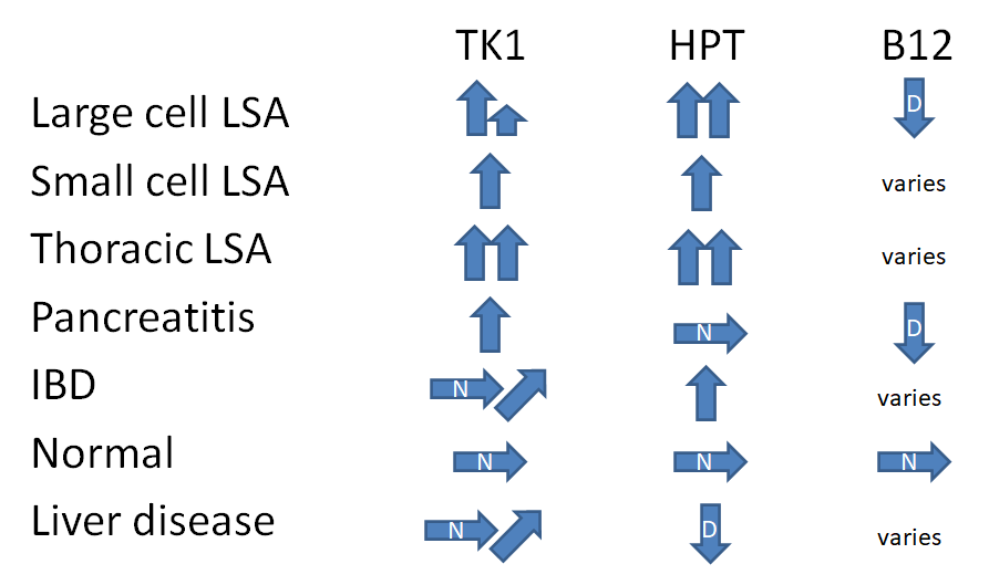
Profiles in the untreated patient
In the untreated patient, the levels of TK1, HPT/CRP, and B12 can indicate different disease states commonly associated with that specific marker profile. Below are some examples of the profiles that are indicated when the resulting markers are increased, decreased, or normal.
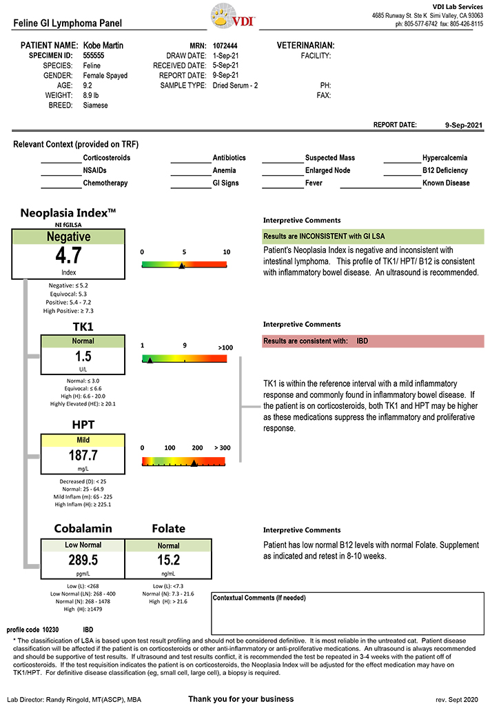
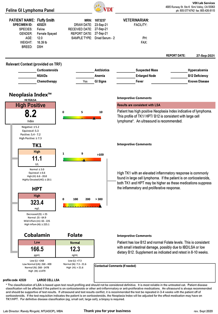
Lab Report Samples
View some samples of actual lab reports by clicking on the images below.