
Cancer Diagnostics
A Cancer Blood Test should be more than yes no maybe.
Get more from your cancer blood tests
MORE…
- Context-sensitive interpretation
- Application-specific cancer panels
- Insight to other concurrent diseases
- Guidance when it’s NOT cancer
- Patient support w/ Test & Treat solutions and supportive tests for the sick animal
- Baseline for monitoring
- VALUE from a single blood test

MORE…
- Patient management
- The BEST tool for monitoring cancer therapy and disease progression
- Minimal sample volume, low cost
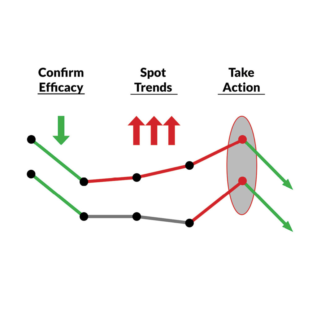
- Identify significant changes, trends, and detect disease recurrence
- Adjust therapy protocol when ineffective
Monitoring That Makes Sense
VDI Cancer Blood Tests
| SERVICE | TEST CODE | NUMBER OF PARAMETERS | SAMPLE VOLUME | INTENDED USE |
|---|---|---|---|---|
| Cancer Panel – General | 201 | 3 | 0.5 mL serum | Initial: ADR or suspicious mass Serial: • Disease progression • Therapy efficacy • Metastasis • Recurrence |
| Cancer Panel – Lymphoma | 201b | 3 | 0.5 mL serum | Initial: Suspected LSA Serial: • Disease progression • Therapy efficacy • Metastasis • Recurrence |
| Pre-Stem Cell Therapy Panel | 205 | 4 | 0.75 mL serum | Initial: Patient evaluation prior to stem cell therapy |
| Pericardial Effusion Panel | 206 | 2 | 0.25 mL serum x2 | Initial: Suspected heart cancer |
| GI Lymphoma Panel | 228 | 5 | 0.5 mL serum | Initial: IBD vs GI LSA Serial: • Disease progression • Therapy efficacy • GI Vitamin status |
| Advanced GI Panel (learn more) | 260 | 12 | 1 mL serum | Initial: IBD, LSA, Pancreatitis, PLE, SIBO, Malabsorption, Dietary Deficiencies Serial: • Reflex to base cancer panel for Cancer/IBD monitoring. • Reflex to GI Vitamins panel for nutritional monitoring |
REFERENCES – Body of Evidence

Boyé, Pierre, et al. “Evaluation of Serum Thymidine Kinase 1 Activity as a Biomarker for Treatment Effectiveness and Prediction of Relapse in Dogs With non‐Hodgkin Lymphoma.” Journal of Veterinary Internal Medicine, vol. 33, no. 4, Wiley, May 2019, pp. 1728–39. https://doi.org/10.1111/jvim.15513.
Euler, Henrik von, Anders B. Öhrvik, et al. “A Non-radiometric Method for Measuring Serum Thymidine Kinase Activity in Malignant Lymphoma in Dogs.” Research in Veterinary Science, vol. 80, no. 1, Feb. 2006, pp. 17–24. https://doi.org/10.1016/j.rvsc.2005.05.001.
Euler, Henrik von. “Monitoring Therapy in Canine Malignant Lymphoma and Leukemia With Serum Thymidine Kinase 1 Activity – Evaluation of a New, Fully Automated Non-radiometric Assay.” International Journal of Oncology, Jan. 1992, https://doi.org/10.3892/ijo_00000175.
Euler, Henrik von, Roland Einarsson, et al. “Serum Thymidine Kinase Activity in Dogs With Malignant Lymphoma: A Potent Marker for Prognosis and Monitoring the Disease.” Journal of Veterinary Internal Medicine, vol. 18, no. 5, Aug. 2004, p. 696. https://doi.org/10.1892/0891-6640(2004)18.
Grobman, M., et al. “Serum Thymidine Kinase 1, Canine-C-Reactive Protein, Haptoglobin, and Vitamin D Concentrations in Dogs With Immune-Mediated Hemolytic Anemia, Thrombocytopenia, and Polyarthropathy.” Journal of Veterinary Internal Medicine, vol. 31, no. 5, Wiley, Aug. 2017, pp. 1430–40. https://doi.org/10.1111/jvim.14787.
Larsdotter, S., et al. “Serum Thymidine Kinase Activity in Clinically Healthy and Diseased Horses: A Potential Marker for Lymphoma.” The Veterinary Journal, vol. 205, no. 2, Elsevier BV, Aug. 2015, pp. 313–16. https://doi.org/10.1016/j.tvjl.2015.01.019.
Meachem, Melissa D., et al. “A Comparative Proteomic Study of Plasma in Feline Pancreatitis and Pancreatic Carcinoma Using 2-dimensional Gel Electrophoresis to Identify Diagnostic Biomarkers: A Pilot Study.” Canadian Journal of Veterinary Research-revue Canadienne De Recherche Veterinaire, vol. 79, no. 3, June 2015, pp. 184–89.
NAKAMURA, Noriko, et al. “Plasma Thymidine Kinase Activity in Dogs With Lymphoma and Leukemia.” Journal of Veterinary Medical Science, vol. 59, no. 10, Japanese Society of Veterinary Science, 1997, pp. 957–60. https://doi.org/10.1292/jvms.59.957.
Selting, K. A., et al. “Serum Thymidine Kinase 1 and C‐reactive Protein as Biomarkers for Screening Clinically Healthy Dogs for Occult Disease.” Veterinary and Comparative Oncology, vol. 13, no. 4, Wiley, July 2013, pp. 373–84. https://doi.org/10.1111/vco.12052.
Selting, K.A., et al. “Thymidine Kinase Type 1 and C‐Reactive Protein Concentrations in Dogs With Spontaneously Occurring Cancer.” Journal of Veterinary Internal Medicine, vol. 30, no. 4, Wiley, May 2016, pp. 1159–66. https://doi.org/10.1111/jvim.13954.
Taylor, Samantha S., et al. “Serum Thymidine Kinase Activity in Clinically Healthy and Diseased Cats: A Potential Biomarker for Lymphoma.” Journal of Feline Medicine and Surgery, vol. 15, no. 2, SAGE Publications, Oct. 2012, pp. 142–47. https://doi.org/10.1177/1098612×12463928.
Thamm, D. H., et al. “Elevated Serum Thymidine Kinase Activity in Canine Splenic Hemangiosarcoma*.” Veterinary and Comparative Oncology, vol. 10, no. 4, Wiley, Oct. 2011, pp. 292–302. https://doi.org/10.1111/j.1476-5829.2011.00298.x.
Cancer Panel
Blood tests for the detection and management of suspected cancer
Cancer Panel
The Cancer panel is dual-marker blood test for diagnosing and managing cancer in a variety of tumor types in dogs and cats. Commonly used as a first-step diagnosis prior to, or in place of biopsy, the cancer panel can give you confidence that neoplasia is present, and if proceeding to more invasive costly workup or referral to specialists is warranted. After diagnosis is made, the Cancer Panel can be used to monitor disease progression or therapy outcomes.
Common Applications:
- Lymphoma
- Abdominal mass
- Solid tumors
- Mast Cell Tumors (grade II or III)
- ADR dog/cat
- Hypercalcemia of malignancy
- Chemotherapy management
- Post mass-removal confirmation
- Metastatic Disease
Not recommended for:
- Cutaneous masses
- Mast Cell Tumors (grade I)
- Brain tumors
Reports Include:
- TK1 result
- C-Reactive Protein (dog) or Haptoglobin (cat) result
- Neoplasia Index (NI)®
- Interpretive Comments
- Monitoring retest recommendations
- Contextual Review (must provide context on requisition form)
Sample Reports:
Cancer Panel – Initial
Cancer Panel – Monitoring
REFERENCES – Body of Evidence
Boyé, Pierre, et al. “Evaluation of Serum Thymidine Kinase 1 Activity as a Biomarker for Treatment Effectiveness and Prediction of Relapse in Dogs With non‐Hodgkin Lymphoma.” Journal of Veterinary Internal Medicine, vol. 33, no. 4, Wiley, May 2019, pp. 1728–39. https://doi.org/10.1111/jvim.15513.
Euler, Henrik von, Anders B. Öhrvik, et al. “A Non-radiometric Method for Measuring Serum Thymidine Kinase Activity in Malignant Lymphoma in Dogs.” Research in Veterinary Science, vol. 80, no. 1, Feb. 2006, pp. 17–24. https://doi.org/10.1016/j.rvsc.2005.05.001.
Euler, Henrik von. “Monitoring Therapy in Canine Malignant Lymphoma and Leukemia With Serum Thymidine Kinase 1 Activity – Evaluation of a New, Fully Automated Non-radiometric Assay.” International Journal of Oncology, Jan. 1992, https://doi.org/10.3892/ijo_00000175.
Euler, Henrik von, Roland Einarsson, et al. “Serum Thymidine Kinase Activity in Dogs With Malignant Lymphoma: A Potent Marker for Prognosis and Monitoring the Disease.” Journal of Veterinary Internal Medicine, vol. 18, no. 5, Aug. 2004, p. 696. https://doi.org/10.1892/0891-6640(2004)18.
Grobman, M., et al. “Serum Thymidine Kinase 1, Canine-C-Reactive Protein, Haptoglobin, and Vitamin D Concentrations in Dogs With Immune-Mediated Hemolytic Anemia, Thrombocytopenia, and Polyarthropathy.” Journal of Veterinary Internal Medicine, vol. 31, no. 5, Wiley, Aug. 2017, pp. 1430–40. https://doi.org/10.1111/jvim.14787.
Larsdotter, S., et al. “Serum Thymidine Kinase Activity in Clinically Healthy and Diseased Horses: A Potential Marker for Lymphoma.” The Veterinary Journal, vol. 205, no. 2, Elsevier BV, Aug. 2015, pp. 313–16. https://doi.org/10.1016/j.tvjl.2015.01.019.
Meachem, Melissa D., et al. “A Comparative Proteomic Study of Plasma in Feline Pancreatitis and Pancreatic Carcinoma Using 2-dimensional Gel Electrophoresis to Identify Diagnostic Biomarkers: A Pilot Study.” Canadian Journal of Veterinary Research-revue Canadienne De Recherche Veterinaire, vol. 79, no. 3, June 2015, pp. 184–89.
NAKAMURA, Noriko, et al. “Plasma Thymidine Kinase Activity in Dogs With Lymphoma and Leukemia.” Journal of Veterinary Medical Science, vol. 59, no. 10, Japanese Society of Veterinary Science, 1997, pp. 957–60. https://doi.org/10.1292/jvms.59.957.
Selting, K. A., et al. “Serum Thymidine Kinase 1 and C‐reactive Protein as Biomarkers for Screening Clinically Healthy Dogs for Occult Disease.” Veterinary and Comparative Oncology, vol. 13, no. 4, Wiley, July 2013, pp. 373–84. https://doi.org/10.1111/vco.12052.
Selting, K.A., et al. “Thymidine Kinase Type 1 and C‐Reactive Protein Concentrations in Dogs With Spontaneously Occurring Cancer.” Journal of Veterinary Internal Medicine, vol. 30, no. 4, Wiley, May 2016, pp. 1159–66. https://doi.org/10.1111/jvim.13954.
Taylor, Samantha S., et al. “Serum Thymidine Kinase Activity in Clinically Healthy and Diseased Cats: A Potential Biomarker for Lymphoma.” Journal of Feline Medicine and Surgery, vol. 15, no. 2, SAGE Publications, Oct. 2012, pp. 142–47. https://doi.org/10.1177/1098612×12463928.
Thamm, D. H., et al. “Elevated Serum Thymidine Kinase Activity in Canine Splenic Hemangiosarcoma*.” Veterinary and Comparative Oncology, vol. 10, no. 4, Wiley, Oct. 2011, pp. 292–302. https://doi.org/10.1111/j.1476-5829.2011.00298.x.
Pericardial Effusion Panel
The Canine Pericardial Effusion Panel uses TK1 from serum and pericardial fluid to determine if the source of the effusion is due to cancer. Neoplastic causes of PCE, particularly hemangiosarcoma, impart a significantly worse prognosis than non-neoplastic etiologies. Ratio of TK1 serum to pericardial fluid is able to rule-out cancer effectively. Contact VDI for more information on the Pericardial Effusion Panel.
GI Lymphoma Panel
Blood tests for the detection and management of suspected GI cancer
GI Lymphoma Panel
The GI Lymphoma Panel is a 4-marker panel used for the diagnosis of GI LSA in the acutely ill cat or dog. Cats are notorious for GI inflammatory disease, and it continues to be one of the harder determinations we see. As GI disease progresses if often times overlaps with the development of Lymphoma – making the differential determination challenging using ultrasound alone. Reluctance and inability to biopsy leaves diagnosis a guess. The GI LSA panel takes the guesswork out.
Applications:
- GI Lymphoma
- Chronic progressive IBD
- ADR cat or dog
- Chemotherapy management
Reports Include:
- TK1 result
- HPT (cat) or CRP (dog) result
- B12/ Folate results
- B12 dosing guidelines
- Neoplasia Index® (NI) GILSA
- Interpretive Comments
- Contextual Review (must provide context on requisition form)
Sample Reports:
REFERENCES – Body of Evidence
Boyé, Pierre, et al. “Evaluation of Serum Thymidine Kinase 1 Activity as a Biomarker for Treatment Effectiveness and Prediction of Relapse in Dogs With non‐Hodgkin Lymphoma.” Journal of Veterinary Internal Medicine, vol. 33, no. 4, Wiley, May 2019, pp. 1728–39. https://doi.org/10.1111/jvim.15513.
Euler, Henrik von, Anders B. Öhrvik, et al. “A Non-radiometric Method for Measuring Serum Thymidine Kinase Activity in Malignant Lymphoma in Dogs.” Research in Veterinary Science, vol. 80, no. 1, Feb. 2006, pp. 17–24. https://doi.org/10.1016/j.rvsc.2005.05.001.
Euler, Henrik von. “Monitoring Therapy in Canine Malignant Lymphoma and Leukemia With Serum Thymidine Kinase 1 Activity – Evaluation of a New, Fully Automated Non-radiometric Assay.” International Journal of Oncology, Jan. 1992, https://doi.org/10.3892/ijo_00000175.
Euler, Henrik von, Roland Einarsson, et al. “Serum Thymidine Kinase Activity in Dogs With Malignant Lymphoma: A Potent Marker for Prognosis and Monitoring the Disease.” Journal of Veterinary Internal Medicine, vol. 18, no. 5, Aug. 2004, p. 696. https://doi.org/10.1892/0891-6640(2004)18.
Grobman, M., et al. “Serum Thymidine Kinase 1, Canine-C-Reactive Protein, Haptoglobin, and Vitamin D Concentrations in Dogs With Immune-Mediated Hemolytic Anemia, Thrombocytopenia, and Polyarthropathy.” Journal of Veterinary Internal Medicine, vol. 31, no. 5, Wiley, Aug. 2017, pp. 1430–40. https://doi.org/10.1111/jvim.14787.
Larsdotter, S., et al. “Serum Thymidine Kinase Activity in Clinically Healthy and Diseased Horses: A Potential Marker for Lymphoma.” The Veterinary Journal, vol. 205, no. 2, Elsevier BV, Aug. 2015, pp. 313–16. https://doi.org/10.1016/j.tvjl.2015.01.019.
Meachem, Melissa D., et al. “A Comparative Proteomic Study of Plasma in Feline Pancreatitis and Pancreatic Carcinoma Using 2-dimensional Gel Electrophoresis to Identify Diagnostic Biomarkers: A Pilot Study.” Canadian Journal of Veterinary Research-revue Canadienne De Recherche Veterinaire, vol. 79, no. 3, June 2015, pp. 184–89.
NAKAMURA, Noriko, et al. “Plasma Thymidine Kinase Activity in Dogs With Lymphoma and Leukemia.” Journal of Veterinary Medical Science, vol. 59, no. 10, Japanese Society of Veterinary Science, 1997, pp. 957–60. https://doi.org/10.1292/jvms.59.957.
Selting, K. A., et al. “Serum Thymidine Kinase 1 and C‐reactive Protein as Biomarkers for Screening Clinically Healthy Dogs for Occult Disease.” Veterinary and Comparative Oncology, vol. 13, no. 4, Wiley, July 2013, pp. 373–84. https://doi.org/10.1111/vco.12052.
Selting, K.A., et al. “Thymidine Kinase Type 1 and C‐Reactive Protein Concentrations in Dogs With Spontaneously Occurring Cancer.” Journal of Veterinary Internal Medicine, vol. 30, no. 4, Wiley, May 2016, pp. 1159–66. https://doi.org/10.1111/jvim.13954.
Taylor, Samantha S., et al. “Serum Thymidine Kinase Activity in Clinically Healthy and Diseased Cats: A Potential Biomarker for Lymphoma.” Journal of Feline Medicine and Surgery, vol. 15, no. 2, SAGE Publications, Oct. 2012, pp. 142–47. https://doi.org/10.1177/1098612×12463928.
Thamm, D. H., et al. “Elevated Serum Thymidine Kinase Activity in Canine Splenic Hemangiosarcoma*.” Veterinary and Comparative Oncology, vol. 10, no. 4, Wiley, Oct. 2011, pp. 292–302. https://doi.org/10.1111/j.1476-5829.2011.00298.x.
Equine Lymphoma
Thymidine Kinase Type 1 – Equine Lymphoma Test
Thymidine Kinase Type 1 (TK1) is a DNA proliferation marker that has been shown in a number of human and animal studies to be highly elevated in Lymphoma. High cellular turnover, especially in aggressive & poorly differentiated lymphoma, gives lymphoma a robust TK1 response that is readily picked up in blood serum. Measurement of TK1 activity can be a rule-in for lymphoma in the suspected horse.